 Colour Perception – The Eye of the Beholder
Seeing the World in glorious colours is central to our lives. Colours shape the way we behave. They affect our mood and our perception. They can influence the way we interact and respond to social and environmental stimuli, whether we are directly aware of it, or through subliminal awareness of our external world. Again, it is one of those things that most people take for granted in everyday life.
Colour perception is all subjective. Colours only exist when three components are present: a viewer, an object, and light.
Around 1665, Isaac Newton discovered that white light splits into its component colours when passing through a dispersive prism. But also, that if those bands of coloured light shine through another prism and re-join, they gather back up and emit a white beam.
Although pure white light is perceived as being colourless, it actually contains all the colours in the visible spectrum. When white light hits an object, it selectively blocks some colours and reflects others.
Only the colours that are reflected, contribute to the viewer’s colour perception.
 Human Colour Perception
The wavelength of light determines what colour we see. From low to high frequency, the perceived colours are: red, orange, yellow, green, cyan, blue, violet.
All the ‘colours of the rainbow‘, as they are often named.
Sufficient differences in frequency give rise to a difference in perceived hue. The just-noticeable difference in wavelength varies from about 1 nm (nanometre = 10-9 m) in the blue/green and yellow wavelengths, to 10 nm and more in the red and blue. (In Psychophysics, this unit is the smallest detectable difference between a starting and secondary level of a particular sensory stimulus.)
Two identical colours can be perceived as being different.
Additionally, our perception of a colour is strongly influenced by the colours surrounding it. Actually, two identical colours can be perceived as being different from one another, if the background colour is different.
Check out these clever optical illusions and you will see exactly what I mean by that:
http://www.echalk.co.uk/amusements/OpticalIllusions/colourPerception/colourPerception.html
Retinal Neurons and the Human Eye
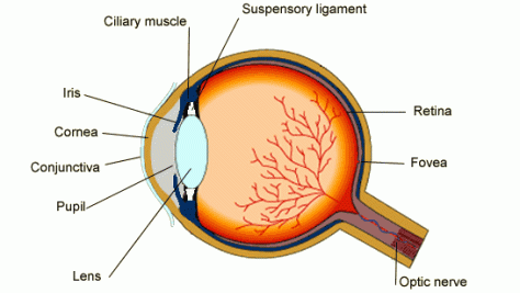
The human eye contains many cells dedicated to making sense of complex external visual stimuli.
Composed of a great number of neurons (photoreceptors. horizontal cells, bipolar cells, amacrine cells and ganglion) that can process visual information, the retina is technically a part of the brain.
Neurons within the retina even project to other parts of the brain, other than the visual cortex, including a specialised area of the hypothalamus controlling body rythms.
The eye senses the visible spectrum using a combination of rod and cone cells gathered on the retina.
In very low light levels, vision is said to be ‘scotopic‘. The light is detected by the rod cells of the retina.
Rods are maximally sensitive to wavelengths near 500 nm, and play little, if any, role in colour vision. Rod cells are more adept at vision under low-light conditions. The rods are more numerous than the cone cells (some 120 millions), and they are also more sensitive than the cones. But they can only sense the intensity of light. They are not sensitive to colour.
In brighter conditions, such as daylight, vision is ‘photopic‘. The light is detected by the cone cells which are responsible for our colour perception.
Cone cells can discern colour, but they function best in bright light. About 6 to 7 million cones provide the eye’s colour sensitivity. They are much more concentrated in the central yellow spot known as the macula.
At the centre of that region is the ‘fovea centralis‘, a 0.3 mm diameter rod-free area with very thin and densely packed cones. This area of the retina provides the clearest, most distinct vision.

The set of possible signals at all three cone cells determines the range of colours we can see with our eyes:
-
S-cones can detect short wavelengths associated with blue colours.
-
M-cones detect medium wavelength in the green range.
-
L-cones detect long (red) wavelengths.
Each of the three standard colour-detecting cones in the retina – blue, green and red – can pick up about 100 different gradations of colour. But the brain can combine those variations exponentially, so that the average person can distinguish about one million different hues.
This diagram illustrates the relative sensitivity of each type of cell for the entire visible spectrum. These curves are known as the ‘tristimulus’ functions.
Colour vision is also an active process that depends as much on the function of the brain, based on previously stored experience, as it does upon the external physical environment.
Colour Variation is in Nature, but what is the Nature of Colour Perception?
The brain determines which colour we see by comparing the combination of inputs received by the three types of cone cells. Therefore, the way we experience colours differs from one individual to another.
Colour blindness occurs when either one photo-pigment is missing, or when it responds maximally to a different wavelength. True colour blindness, or achromatopsia, is a rare, hereditary, sex-linked condition, associated with the X-chromosome.
About 30 in a million people do not see any colours, and perceive the world as if in black and white. Neil Harbisson has achromatopsic vision. He deals with his condition in a creative fashion, using a clever little device called the ‘eyeborg‘, which lets him listen to colours.
1 in every 12 men, and 1 in every 200 women, are affected by some form of colour deficiency.
Dichromacy may occur if there is a complete loss of one type of cones. Anomalous trichromacy, a shift in spectral sensitivity of one type of cones (affecting L-cones and M-cones), is a rather common form of colour deficiency. Typically, it manifests as a confusion between red and green, which occurs due to a variation in the cone pigment caused by a difference in amino-acid sequences.

At the opposite end of the scale, a tetrachromat has another type of cone, between the red and green (somewhere in the orange range). For such an individual, statistically more likely to be female, 100 shades would theoretically enable her to see 100 million different colours.
Colour deficiency is often diagnosed using the Ishihara Colour Test, which consists of a series of pictures of coloured spots, to assess red-green colour deficiencies.
Mapping the Human Brain with Retinal Neurons
The human brain has 100 billion neurons, connected to each other in networks (or synapses) that allow us to interpret the world around us, plan for the future, and control our actions and movements. Along with other neuroscientists from MIT, Sebastian Seung wants to map those networks, and create a wiring diagram of the brain that could help scientists learn how we each become our unique selves.
Retinal neurons, being part of the brain, provide a more approachable starting point. By mapping all of the neurons on the retina of a mouse, a mere 117 μm (or micrometer = 10-6 m) by 80 μm little patch of tissue, MIT researchers were able to classify most of the neurons they found, based on their patterns of wiring. The team also identified a new type of retinal cell that had not been seen before.
All in all, the retina is now estimated to contain 50 to 100 different types of neurons, which have until now remained exhaustively uncharacterised.
Read more on MIT Article ‘Making Connections in the Eye’…
Earlier in 2013, President Obama announced funding of $100 million for a new initiative into research and technologies that would allow for a better understanding of the human brain. The Brain Initiative has been compared to the Human Genome Project (HGV), because it is directed at a problem that has seemed insoluble until now: recording and mapping brain circuits in action, in an effort to “show how millions of brain cells interact”.
The effort will require neuroscientists to develop new tools and, will perhaps ultimately lead to progress being made in the treatment of diseases like Alzheimer’s, epilepsy, stroke and brain trauma. It will involve both government agencies and private institutions.
But if you’ve ever wondered what it would feel like to see the World through someone else’s eyes, future research may soon provide the answer…

